Protective mechanism of the inner ear from traumatic noise exposure actuated by conditioning with heat stress
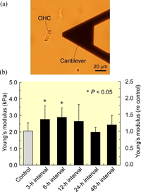
Fig. 1. Measurement of Young's moduli of outer hair cells (OHCs) in mice by atomic force microscopy. (a) Schema of the measurement. (b) Changes in Young's moduli of OHCs before and after heat stress.
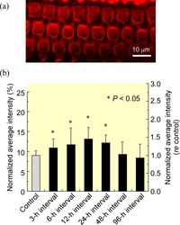
Fig. 2. Observation of filamentous actin of outer hair cells in mice by confocal laser scanning microscopy. (a) Fluorescence image of filamentous actin at the lateral wall of OHCs. (b) Changes in the amount of filamentous actin of OHCs before and after heat stress.
High hearing sensitivity of mammals is made possible by cochlear amplification due to the motility of outer hair cells (OHCs). However, since OHCs are nonrenewable, overexposure to intense sound damages them and causes the loss of cochlear amplification, resulting in permanent hearing loss.
Recently, it has been found that the OHCs can be protected from such traumatic exposure by prior sublethal conditioning with heat stress. A possible reason for this protective mechanism is modification of the cell stiffness which reduces the mechanical damage of cells induced by traumatic exposure. However, it is unclear whether the stiffness of the cell increases or decreases by such modification.
In this study, therefore, the mechanical properties and the amount of filamentous actin (F-actin) of the OHCs in mice with or without whole-body heat stress were measured by atomic force microscopy (AFM) and confocal laser scanning microscopy (CLSM), respectively.
The conditioning by heat stress caused an increase in Young's modulus (Fig. 1) and F-actin (Fig. 2) of the OHCs in mice 3-12 h after heat stress. These results suggest that OHCs exposed to prior heat stress possibly experience less strain when they are exposed to loud noise, resulting in protection from traumatic noise exposure.
- Kitsunai Y†, Yoshida N†, Murakoshi M†, Iida K, Kumano S, Kobayashi T, Wada H. Effects of heat stress on filamentous actin and prestin of outer hair cells in mice. Brain Res 1177: 47-58, 2007. †These authors contributed equally to this work.
- Murakoshi M, Yoshida N, Kitsunai Y, Iida K, Kumano S, Suzuki T, Kobayashi T, Wada H. Effects of heat stress on Young's modulus of outer hair cells in mice. Brain Res 1107: 121-130, 2006.
- Murakoshi M, Yoshida N, Iida K, Kumano S, Kobayashi T, Wada H. Local mechanical properties of mouse outer hair cells. atomic force microscopic study. Auris Nasus Larynx 33: 149-157, 2006.