Three rows of outer hair cells are required for cochlear amplification

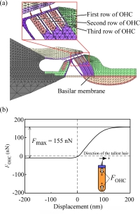
Fig. 1. Finite model used in the present study. (a) Model of the OC. (b) Estimated force generated by the OHC.

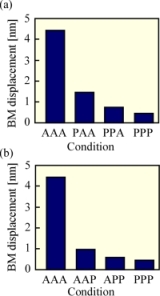
Fig. 2. Change in the BM displacement caused by functional loss of OHCs at 60 dB SPL. (a) Functional loss from the innermost row of OHC. (b) Functional loss from the outermost row of OHC. A and P represent active and passive, respectively.
The process known as cochlear amplification is realized by coordinated movement of the outer hair cells (OHCs) in response to changes in their membrane potential. In this process, the displacement amplitude of the basilar membrane (BM) is thought to be increased, thereby leading to the high sensitivity, wide dynamic range and sharp frequency selectivity of our hearing. Unfortunately, however, OHCs are vulnerable to noise exposure, ototoxic acid, aging and so on.
Previous studies have shown that exposure to intense noise causes functional loss of OHCs from the innermost row (i.e., close to the modiolus) to the outermost row (i.e., close to the cochlear wall). On the contrary, by other traumatic stimuli such as ototoxic acid, aging and ischemia, such loss of OHCs has been reported to occur from the outermost row toward the innermost row. However, how the cochlear amplification changes when coordinated movement of OHCs is impaired, that is when the OHCs in one, two or all three rows have become dysfunctional, remains unclear.
In the present study, therefore, a finite element (FE) model of the gerbil cochlea, which takes the motility of OHCs into account, was developed based on our previous FE model (Fig. 1). Using this model, changes in the displacement amplitude of the BM due to the functional loss of OHCs in one, two or all three rows were investigated and the effects of incoordination of the three rows of OHCs on cochlear amplification were estimated.
Results showed that the displacement amplitude of the BM significantly decreased when either the innermost row or the outermost row of OHCs lost its function and that the outermost OHCs possibly has larger effect on the displacement amplitude of the BM than the innermost OHCs. These results suggest that all three rows of OHCs are required for cochlear amplification (Fig. 2).
This study was conducted in collaboration with Professor Hiroshi Wada, Tohoku Bunka Gakuen University, Sendai, Japan.