Imaging by atomic force microscopy of the motor protein prestin
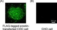
Fig. 1. Fluorescence images of prestin labeling in the prestin-transfected and untransfected CHO cells. (A) Prestin-transfected CHO cell. (B) Untransfected CHO cell. Prestin was expressed in the plasma membranes of the prestin-transfected CHO cells.
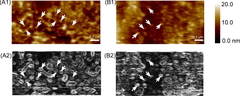
Fig. 2. AFM images of the membranes of the CHO cells. (A1) Original AFM image of a prestin-transfected CHO cell. (A2) Differential AFM image of A1. (B1) Original AFM image of an untransfected CHO cell. (B2) Differential AFM image of B1. Particle-like structures can be seen in these AFM images (arrows).
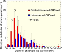
Fig. 3. Frequency distribution of the observed particle-like structures in the CHO plasma membrane. The density of the particle-like structures 8 - 12 nm in diameter in the prestin-transfected CHO cells is higher than that in the untransfected CHO cells with statistical significance.
The motility of outer hair cells (OHCs) is thought to be based on the motor protein prestin. However, structure of prestin has not yet been clarified.
In the present study, the plasma membranes of prestin-transfected and untransfected Chinese hamster ovary (CHO) cells were observed using an atomic force microscope (AFM) to visualize prestin.
Figure 1 shows the fluorescence images of prestin labeling in the prestin-transfected and untransfected CHO cells. Prestin was expressed in the plasma membranes of the prestin-transfected CHO cells. Figure 2 shows the AFM images of the plasma membranes of the prestin-transfected and untransfected CHO cells. Particle-like structures were observed in the plasma membranes of both cells. Analysis of the size of the observed structures was therefore performed.
Based on the molecular weight of prestin (~82 kDa), its diameter is thought to be ~7 nm if its shape is assumed to be cubical. Previous morphological studies by electron microscopy have found the lateral wall of OHCs to be covered with 7 - 25-nm particles. When the ratio of the length of the minor axis to that of the major axis was 0.5 - 1.0, the structure was regarded as being a particle-like structure. When the sizes of the particle-like structures in the plasma membranes were 8 - 10 nm and 10 - 12 nm, the differences of their densities between the prestin-transfected CHO cells and the untransfected CHO cells were statistically significant (Fig. 3). Since the difference between these two types of cells is due to the existence of prestin, approximately 75% of the particle-like structures with a diameter of 8 - 12 nm in the prestin-transfected CHO cells are possibly constituted of prestin.